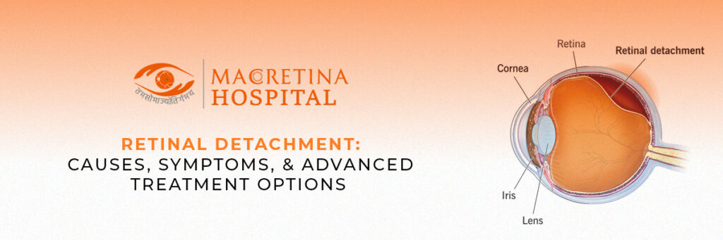
If you leave retinal detachment untreated, it will become an eye condition that will cause a person permanent loss of vision. The retina, which is the sensitive layer situated in the back of the eye and which works by sending signals to the brain to make vision possible, pulls or separates itself from the underlying tissue. So, in a way, this disruption will prevent the normal functioning of the retina, which may impair sight. In fact, it can cause total blindness in certain cases. Early diagnosis and treatment are essential for preventing long-term damage to the vision.
If you happen to notice any symptoms like the above, don’t forget to take prompt medical attention to a reputable hospital or eye care center such as Macretina Hospital. Now more than ever, the advent of advanced modern diagnostic equipment coupled with surgical techniques makes it very manageable. Read on as we discuss the causes, symptoms, risk factors, and advanced treatment options available for this condition. Consulting with experts in the finest eye clinic Indore can ensure you get the best care to preserve your vision.
What is Retinal Detachment?
Retinal detachment happens when the retina becomes lifted or pulled away from its normal position. Considered a medical emergency that requires immediate attention to prevent vision loss, retinal detachment could have myriad causes, including retinal tears, fluid accumulation, or scarring.
Types of Retinal Detachment
Retinal detachments can be mainly classified into three types:
- Rhegmatogenous retinal detachment: This refers to the most common type of retinal detachment induced by a tear or hole in the retina. Fluid from the vitreous cavity (gel-like substance inside the eye) seeps through the tear, causing detachment of the retina.
- Tractional Retinal Detachment: This detachment occurs when scar tissue or fibrous bands pull the retina away from the back of the eye. This type of retinal detachment is mostly seen in people suffering from diabetic retinopathy or other vascular diseases.
- Exudative retinal detachment: This type occurs when fluid accumulates beneath the retina without any tears or holes. This detachment is often associated with an inflammatory disease, a tumor, or systemic conditions.
Causes and Risk Factors
Common Causes of Retinal Detachment
Retinal detachment is typically due to one of several factors:
- Aging: Over time, the eye’s vitreous gel shrinks within the eye, pulling away from the retina, and occasionally creating tears or hole.
- High Myopia or Severe Nearsightedness: Longer eyeballs in patients with high myopia predispose them to retinal thinning and subsequent detachment.
- Eye Traumas: Blunt force injury or penetrating trauma will cause tears in the retina leading to retinal detachment.
- Former Eye Surgery: Such as, cataract surgery increases the risk that any repair or procedure on the eye will result in the use of retina detachment.
- Diabetes: The penetration of the organ has been observed to lead to abnormal growth of blood vessels and scarring, pulling with it the retina.
- Family History: A person is likely to get closer to this retinal attachment if the family has a genetic tendency.
- Inflammatory Eye Conditions: These include uveitis that leads to fluid accumulation beneath the retina, resulting in detachment.
Retinal Detachment Symptoms
Identifying symptoms of retinal detachment at the initial stages can prevent visual impairment from becoming permanent. The common warning signs include:
- A sudden increase in floaters: These are tiny dark specks or cobweb-like shapes drifting in your vision.
- Flashes of light: Sudden and bright flashes, particularly when viewed in peripheral vision.
- Shadow or curtain effect: A dark shadow being cast or a curtain moving across the field of vision.
- Blurred vision: Blurriness or some unexplained loss of sharpness in vision.
- Peripheral vision loss: Gradual loss of side vision.
If you have any of these symptoms, immediately contact an eye specialist at the best eye clinic in Indore to prevent further complications.
Diagnosis and Eye Examination
It takes a comprehensive eye examination to confirm retinal detachment. Some of the common diagnostic tests include:
- Dilated Eye Exam: The doctor will use special eye drops to widen the pupils and examine the retina for any tears or detachment.
- Ultrasound Imaging (B-scan): This test is used for detection of retinal detachment in a case where bleeding or cataract completely obscures the retina.
- OCT: This is high-resolution imaging that gives detailed cross-sectional views of the retina.
- FFA: The blood flow abnormalities in the retina will be identified by injecting a dye into the bloodstream.
Early detection is vital for vision conservation. Contact one of the best retina surgeons in Indore for an accurate diagnosis and an appropriate treatment plan.
Better Treatment Options for Patients Suffering from Retinal Detachment
Depending on the stage of detachment, there are several treatment options available.
Non-Surgical Treatments
- Laser Photocoagulation: The laser is used to seal small retinal tears so that fluid cannot pass through.
- Cryotherapy: An external freezing probe is applied to the eye to create scar tissue that serves to help seal the retina.
Surgical Treatments
- Pneumatic Retinopexy: A gas bubble is injected into the eye to push the retina back in place. The patient maintains a specific head position so that the gas bubble does not move out of place.
- Scleral Buckling Surgery: A silicone sponge is placed around the eye to neutralize the pulling forces and assist in the reattachment of the retina.
- Vitrectomy: The vitreous gel is extracted and replaced with gas or silicone oil to push the retina back into position.
Recovery and Aftercare
Proper aftercare should be given to these patients for a successful recovery. Key components are:
- Follow all post-surgical medications and instructions including restrictions against heavy lifting or vigorous activities.
- Head Position: In comparison, lying in the proper head position is very important if gas bubble surgery was done.
- Regular follow-ups: To track retinal health on a scheduled basis.
- Lifestyle and Diet: Antioxidants as well as omega-3 fatty acids and vitamins A, C, and E are important for healing.
- Avoiding exposure to bright lights: To spare the eyes from any excess light, it is advisable to underpin wearing UV-protective glasses.
Protective Steps for Your Retinal Health
Not all cases of retinal detachment can be prevented, but some measures can reduce the risk.
- Regular monitoring of eyes: In particular, those with high myopia or a family background of retinal problems.
- Good control of diabetes and hypertension: Keeping these conditions under control decreases the retinal complications.
- Reduced chances of eye injury: Wearing protective eyewear while participating in sports or activities of high risk.
- Identifying early symptom: To be checked immediately when the patient sees floaters, flashes, or loss of vision.
Summary
Retinal detachment is a severe eye emergency, and any delay in treatment can result in permanent loss of vision. However, timely diagnosis and prompt treatment considerably improve prognosis. Therefore, in the event of any such symptoms, visiting a skilled retinal surgeon in Indore is essential for timely intervention.
Expert care at Macretina Hospital offers advanced diagnostic equipment and retinal treatment modalities. With appropriate treatment, retinal detachment can be appropriately managed so as to maintain healthy vision for life.