What is Macular Degeneration?
The macula is the central area of the retina where reading vision is located. The retina is the light sensitive inner lining in the back of the eye that is like the film in a camera. Anatomically, the macula is located in the straight ahead position of the eye and is at the center of the retina. If the retina were a target, the macula would be the bull’s eye. Macular degeneration causes a person’s vision to become blurred, distorted, and/or dark in the straight- ahead direction. While macular degeneration rarely causes loss of all vision, it can make daily activities such as reading or driving difficult or impossible.
What Causes Age-related Macular Degeneration?
Macular degeneration is almost always seen in older people, although in a few families it may start earlier. The prevalence of macular degeneration increases as the population gets older. There is a definite genetic predisposition as shown by studies comparing identical twins and fratemal twins. Other factors that have an effect on the severity of the disease include high-blood pressure and cigarette smoking, It is now the most common cause of legal blindness in retirement-age people in the developed countries and India will follow the same statistics! There are two main forms of macular degeneration: dry and wet.
What is the difference between Wet & Dry Macular Degeneration?
During normal aging, yellowish deposits, called drusens, form under the retina, which is the light-sensitive layer of tissue at the back of the eye that provides clear, sharp images As drusens increase in size and number, they can interfere with proper functioning of the retina, damaging or killing the light-sensitive cells of the macula.
Because the macula’s light-sensitive cells provide the ability to have sharp, detailed vision, the results can be blurring of central vision and a devastating impact on the ability to enjoy activities of daily life, such as reading, driving, or even recognizing the face of a friend or family member. This form of age-related macular degeneration is called dry AMD. Dry AMD can be a precursor to wet AMD.
Wet AMD occurs when abnormal blood vessels behind the retina start to grow under the macula. These blood vessels often leak blood and fluid, damaging or killing light-sensitive cells-loss of vision occurs quickly.
Although approximately 80 percent of patients with age-related macular degeneration have dry AMD, wet AMD is responsible for 80 to 90 percent of severe loss of vision or legal blindness associated with this disease. Symptoms
• Need for brighter light when reading
• Difficulty adapting to low light levels
• Increased blurriness of printed words
• Decrease in brightness of colors
• Blurred spot in the center of the field of vision
• Blank or black spot in the field of vision (spot will start small and grow over time, possibly leading to blindness
• Abrupt decline in central vision
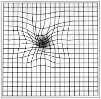
• Visual distortions, such as straight lines appearing wavy, or objects appearing larger or smaller than they are
• Well-defined blind spot in the center of vision.
It is important to pay close attention to any decline in central vision-both near and distant. if you notice any of these signs or symptoms, schedule an examination with a retina specialist. Risk factors
• Cigarette smoking
• Obesity
• Hypertension (high blood pressure)
• Excessive sun exposure
• Diet deficient in fruits and vegetables
Most people who suffer from macular degeneration have the “dry” form. Dry macular degeneration involves changes in the pigment layer of the retina in the back of the eye and the accumulation of yellow deposits called drusen. More light may be required for reading, and it may take longer to adapt to a darker room. Most people maintain the ability to read and drive, but occasionally dry macular degeneration can cause legal blindness. Wet macular degeneration accounts for about 10% of all cases. Wet macular degeneration is the more severe form of the disease. People with wet macular degeneration develop abnormal leaking blood vessels and membranes under the macula. The size, location and type of the leaking blood vessels determine the proper treatment of the disease. With wet macular degeneration, vision loss is often rapid and severe. The wet form can be further classified into classic, occult, polypoid, and retinal angiomatous proliferation (RAP).
What are the Symptoms of Wet Macular Degeneration?
Dry macular degeneration is usually a slow process and is hardly noticeable in its early stages. It is important to detect the transition from dry to wet macular degeneration as soon as possible to get the best effect from vision saving treatments. While symptoms may vary from person to person, there are several ways to detect macular degeneration. These methods work for both wet and dry macular degeneration but a recent onset or worsening of symptoms suggests wet macular degeneration. Common symptoms include:
1) dark areas or holes in the center of your vision,
2) letters or words begin to look blurry when reading and
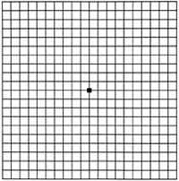
3) straight lines appear distorted or wavy. One simple way to check for macular degeneration is to use the Amsler grid.

How is Macular Degeneration Diagnosed?
Because the early symptoms of wet macular degeneration usually occur in one eye at a time, the good vision in the other eye may cover-up the symptoms of the eye with wet macular degeneration. Many people don’t realize they have the disease until their visual loss is already severe. However, by using the Amsler grid frequently to test your eyes and by testing each eye separately, you can spot the disease early in its course, Regular eye exams also detect early signs of the disease. If you or Dr Pratik suspect macular degeneration, he can use a microscope and ophthalmoscope to get a detailed view of your macula, Another method to detect abnormal blood vessels in the retina is a special series of eye photographs, called a fluorescein angiogram. Optical coherence tomography is a laser scan that demonstrates fluid accumulation in wet macular degeneration.
What is the Treatment for AMD?
The risk of worsening AMD may be reduced by reducing risk factors through maintaining a healthy life style, for example, quitting smoking, eating lots of fruits and vegetables, and exercising regularly. Dry AMD. The Age-Related Eye Disease Study (AREDS) showed that high-dose anti-oxidant vitamins can reduce the risk of progression of dry AMD in some patients. Your doctor can tell you whether these vitamins are appropriate for you. Wet AMD. Many patients with wet AMD can be treated with drugs, such as Lucentis, Accentrix, Pagenax, Eylea, Vabysmo, Razumab , Avastin, etc that are injected directly into the eye to prevent the growth of new blood vessels. Some patients are treated with laser therapy. With laser photocoagulation, a beam of light is used to seal the abnormal blood vessels and prevent leaking. With photodynamic therapy (PDT), a low energy laser is used to activate a lightsensitive drug that is in jected into a vein and travels to abnormal blood vessels in the macula. The light activated drug then destroys the abnormal vessels. Your doctor can determine which, if any. treatment is appropriate for you. In some cases, a combination of treatments may be used.
What if vision is reduced despite the best treatment?
Low vision devices can often greatly improve visual performance in people that are considered legally blind. The two major classes of low vision devices are:
1) Lens Magnification combined with a brighter light
2) Closed Circuit Television (CCTV) with electronic magnification and image intensification
Diagnostic tests
Dr Pratik Mahajan often advices
Copyright@ Macretina Hospital 2026 || All Right Reserved