Eyes are among the delicate organs of the human body and early detection of any problems is crucial to having a good vision. Nowadays, medical imaging devices enable retinal diseases to be diagnosed with more accuracy than previously. The two most important diagnostic tests used in ophthalmology are Fundus Photography and Fluorescein Angiography (FFA). These imaging techniques help specialists in diagnosing and managing a number of retinal disorders together with timely intervention into the case and, therefore, better treatment outcomes.
This article highlights the essence of Fundus Photography and FFA in diagnosis of retinal diseases, how they function, their advantages, and the need to visit a retina specialist in Indore for proper eye care.
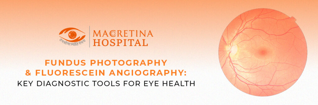
Understanding Fundus Photography
Fundus Photography is an imaging non-invasive technique used to obtain high-resolution images of the interior surface of the eye, namely retina, optic disc, macula, vascular system, so as to monitor and diagnose different retinal and optic diseases.
How does Fundus Photography Work?
A photography session is made possible through a special camera that is fitted with a low-power microscope and applies appropriately to get very detailed images of the interior of the eye. Basically, the patient gazes through at a fixation target while the pictures of the retina are taken. In some cases, dilating drops are also used for clarity of pictures.
Uses of Fundus Photography
Fundus Photography is important for detecting and monitoring a variety of eye diseases. They include among others:
- Diabetic Retinopathy – The detection of retinal blood vessel damage because of the disease.
- Glaucoma – Monitors usually with optic nerve head changes.
- Aging Macular Degeneration (AMD) – Uses for the tracking of changing states of the macula and the development of new blood vessels.
- Hypertensive Retinopathy – Damage from high blood pressure is usually detected.
- Retinal Detachment – Was helpful in detecting areas either torn away from the retina or detached sections of that retina.
Understanding fluorescein angiography (FA)
Fluorescein angiography (FA) is a more intense imaging modality, which involves the injection of a fluorescent dye into the retinal case, revealing the blood flows within the retinal structure. The abnormalities in both retinal and choroidal blood vessels are reported by this method.
How does fluorescein angiography work?
A fluorescent dye is injected into a vein—the arm usually. The dye travels through the bloodstream and reaches the blood vessels of the retina. An image of the retina taken as the dye circulates would capture any abnormal blood vessels or leakage.
Uses of fluorescein angiography
Typically, FFA is used in the diagnosis of:
- Diabetic Retinopathy – Leakage and abnormal growth from blood vessels are detected.
- Macular Edema – Assesses the accumulation of fluid in the macula.
- Retinal-vein occlusion – detects blockages of blood vessels or their damage.
- Choroidal Neovascularization – Diagnosis of neovascularisation (growth of new blood vessels under the retina).
- Uveitis – Identifies changes in vascular lesions attributed to inflammation.
Importance of Fundus Photography and FFA
Both modalities are crucial in the detection of disease in its early stages among other things. Other merits include:
1. Early Diagnosis of Retinal Disorders
Detection of lesions is done before the manifestation of symptoms, enabling early diagnosis and treatment with better outcomes.
2. Non-Invasive and Painless
It was completely non-invasive, although FFA involved only the subcutaneous injection, thus establishing its reliability in diagnosis.
3. Monitoring Disease Progression
Chronic conditions affecting the eyes are amenable to periodic imaging for monitoring disease progress and effectiveness of treatment.
4. A More Precise Treatment Outline
High-resolution images allow ophthalmologists to establish accurate treatment outlines for the better management of retinal ailments.
Who needs to take these tests?
Fundus Photography and FFA are recommended for people who have:
- Diabetes or high blood pressure
- A family history of retinal diseases
- Unexplained vision changes or blurriness
- Previously performed eye surgeries or trauma
- Chronic conditions affecting the eye such as glaucoma or AMD
Consulting a retina specialist in Indore would help with determining whether such tests were necessary for any vision abnormalities.
Risks and Precautions
Fundus Photography has no risks attached to it, while the few that are associated with Fluorescein Angiography include the following:
- Yellow temporary staining of the skin and urine due to dye elimination
- Mild nausea or dizziness post-injection
- Rare allergic reactions to the dye
For safety purposes, the patient should inform his physician regarding allergies or existing health conditions before undergoing FFA.
The importance of Routine Retinal Exam
Vision loss or even early detection of disease is possible through use of advanced diagnostic tools during eye examination. This is especially important in people prone to higher risks. They include persons with diabetes, high blood pressure, and a family history of eye conditions.
Conclusion
Fundus Photography and fluorescein angiography are very significant in the diagnosis and management of retinal diseases. As it provides very specific information concerning the retina’s health, an early detection of problems, and timely treatment can be rendered by ophthalmologists. If you are facing any problems in vision or routine eye checkups, visiting a retina specialist in Indore at best could do justice to the case. Keeping up with regular eye exams and advanced diagnostic tests will help preserve your vision and maintain eye health overall.